


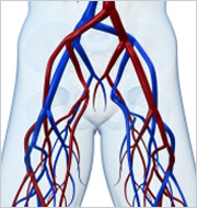
 Renal artery ultrasound is a test that shows the renal arteries, the arteries that carry blood to the kidney. These arteries may narrow or become blocked and this may result in kidney failure or high blood pressure (hypertension). Ultrasound waves—the same ones used in imaging the fetus in a pregnant woman—are used to make an image of the artery. The speed of blood flow through the arteries is measured and determines the degree of narrowing of the artery. Imaging of the renal arteries can be extremely difficult and this test most often is performed in the morning on an empty stomach.
What patients require an renal artery ultrasound?
Doctors will request an renal artery ultrasound on patients who have early signs of kidney failure or blood pressure is difficult to control despite multiple medications.
What happens during an renal artery ultrasound?
You will be asked to lie down on an examination table. The technician (or physician) will place a clear gel on your abdomen. The gel is simply a lubricant that allows the transducer (a device that both puts out and detects ultrasound signals) to slide around easily on your skin. When the transducer is placed against the skin, an image of the artery is shown on a video screen. The renal arteries are identified and a measurement will be made of the speed of blood flow through the artery. This test is typically not performed during the screening program, but patients who may require the study are identified and are set up for the study at a later date.
What are the risks of renal artery ultrasound?
Since the procedure is done without entering the body and does not use dyes or x-rays, there is no risk or pain involved in having a renal artery ultrasound.
How does a renal artery ultrasound work?
The transducer emits high-frequency, ultrasound waves that pass into the body and bounce off the abdominal aorta. The sound waves are reflected differently by different parts of the body. The transducer detects the different reflections of the sound waves, which are then measured and converted by a computer into live pictures of the artery.


