


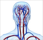
 Carotid artery duplex is a test that shows the carotid arteries (vessels in the neck that provide blood flow to the brain), as well as how much blood flows and how fast it travels through them. Ultrasound waves—the same ones used in imaging the fetus in a pregnant woman—are used to make an image of the arteries. This image can be used to find out if there is an abnormality or blockage of the carotid arteries that could lead to stroke. Minor blockage or narrowing in the carotid artery is common in the elderly but if the degree of narrowing exceeds 50% the risk of stroke increases.
What patients require a carotid artery duplex?
Doctors will request a carotid ultrasound on patients who have either had a stroke, TIA (or mini-stroke) or who might be at high risk for a stroke. These patients include those who have vascular risk factors such as smoking, high blood pressure, diabetes, elevated lipids, or those with a history of coronary artery disease or peripheral vascular disease. Most of the patients who have severe carotid artery disease do not know they have the problem until a stroke occurs, that is why screening programs have developed that include carotid ultrasound.
What happens during a carotid artery duplex?
You will be asked to lie down on an examination table. The technician (or physician) will place a clear gel on the area of the neck where the carotid artery is located. The gel is simply a lubricant that allows the transducer (a device that both puts out and detects ultrasound signals) to slide around easily on your skin. When the transducer is placed against the skin, an image of the artery is shown on a video screen. To view the arteries from many different angles, your doctor will re-position the transducer several times. Because blood is flowing through the artery, a sound similar to your heartbeat will be heard. The procedure is repeated for the carotid artery on the other side of the neck. A carotid ultrasound screening usually only takes a few minutes, the formal examination may take 15 to 30 minutes to complete.
What are the risks of carotid artery duplex?
Since the procedure is done without entering the body and does not use dyes or x-rays, there is no risk or pain involved in having a carotid ultrasound.
How does a carotid artery duplex work?
The transducer emits high-frequency, ultrasound waves that pass into the body and bounce off the carotid arteries and the red blood cells moving through them. The sound waves are reflected differently by different parts of the body. The transducer detects the different reflections of the sound waves, which are then measured and converted by a computer into live pictures of the arteries and the blood flow.


