


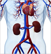
 Abdominal aortic ultrasound is a test that shows the abdominal aorta, the large blood vessel that carries blood through the abdomen. This artery may enlarge or dilate and develop into an aneurysm. Ultrasound waves—the same ones used in imaging the fetus in a pregnant woman—are used to make an image of the artery. This image can be used to find out if there is an enlargement of the artery. If the artery enlarges more than one and one-half times the size of the normal artery, it is considered an aneurysm.
What patients require an abdominal aortic ultrasound?
Doctors will request an abdominal aortic ultrasound on patients who have a pulsatile mass in their abdomen or those patients that have a family history of aneurysms.
What happens during an abdominal aortic ultrasound?
You will be asked to lie down on an examination table. The technician (or physician) will place a clear gel on your abdomen. The gel is simply a lubricant that allows the transducer (a device that both puts out and detects ultrasound signals) to slide around easily on your skin. When the transducer is placed against the skin, an image of the artery is shown on a video screen. The aorta is identified and a measurement will be made of the diameter of the artery. An abdominal aortic ultrasound screening usually only takes a few minutes, the formal examination may take 15 to 30 minutes to complete.
What are the risks of abdominal aortic ultrasound?
Since the procedure is done without entering the body and does not use dyes or x-rays, there is no risk or pain involved in having an abdominal aortic ultrasound.
How does an abdominal aortic ultrasound work?
The transducer emits high-frequency, ultrasound waves that pass into the body and bounce off the abdominal aorta. The sound waves are reflected differently by different parts of the body. The transducer detects the different reflections of the sound waves, which are then measured and converted by a computer into live pictures of the artery.


